
-
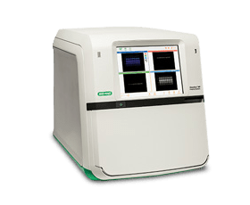
ChemiDoc Imaging Systems
ChemiDoc Imagers offer best-in-class performance with ease of use for visible light (RGB), far/near infrared (FR/NIR) fluorescence, chemiluminescence detection, and stain-free imaging for all western blotting and general gel documentation applications.
-
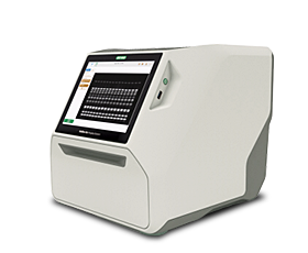
GelDoc Go Gel Imaging System
The GelDoc Go Imaging System is a compact benchtop imaging solution designed for ease of use. Acquire high-resolution, publication-quality images of both nucleic acid and protein gels.
-
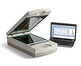
GS-900 Calibrated Densitometer
Achieve fast, reproducible imaging of gels, blots, and film with the GS-900 Calibrated Densitometer. Quantitate proteins across a wide dynamic range using transmissive and reflective imaging.
Gel Documentation Systems from Bio-Rad
Bio-Rad offers a comprehensive range of imaging systems for the detection, quantitation, and analysis of proteins and nucleic acids in gels and on membranes. These gel documentation systems have advanced software, providing rapid automated data acquisition, analysis, and validation. Our gel imaging systems can be used for detection and automated data analysis with all common modes of protein and nucleic acid staining and labeling: colorimetric, fluorescent, chemiluminescent, and radioisotopic.
Most Bio-Rad gel documentation systems also feature our unique stain-free imaging technology, allowing rapid visualization of proteins on gels and blots without staining and destaining (Bio-Rad's TGX Stain-Free or Criterion Stain Free Precast Gels, or TGX Stain-Free FastCast Acrylamide Solutions, are also required).
Gel Documentation System Features
The choice of a gel documentation system depends on both your current and anticipated future needs. Bio-Rad's gel documentation systems accommodate a wide range of samples and support different modes of detection. What features might you need in a gel documentation system?
Modular design and flexible options allow you to customize a system for your specific requirements. Additionally, as your needs change and expand, our gel documentation systems have the flexibility to accommodate different sample types and detection modes.
Multiple modes of data analysis and output provide ease of data evaluation, customized reports, sharing of data, different types of output, and generation of publication-quality images. For analyses that are often repeated, automation of the specific data acquisition, analysis, and output modes can save time and reduce errors. Specific software modules are also offered to satisfy 21 CFR Part 11 regulatory requirements.
Signal accumulation mode (SAM) is the collection of chemiluminescence data over time, allowing the measurement of chemiluminescence as it develops. SAM enables the capture and analysis of a time course of the development of chemiluminescent signals, unlike film-based systems, where an image is captured at a single arbitrary time point. In gel imaging systems with SAM capability, the system determines the best exposure time and captured image. SAM provides fast, optimal results for ECL and other chemiluminescence-based protocols.
Multifluorescent western blot detection allows the simultaneous detection of more than one protein on a blot. This permits the detection of multiple proteins in a single sample, without requiring loading the same sample in multiple wells or stripping and reprobing the blot. A gel imaging system that is capable of multifluorescent detection increases accuracy because there is no error due to inaccuracy in replicate loading or the loss of protein that occurs during stripping a blot for reprobing. In addition to increased accuracy, multifluorescent detection saves time, and fewer reagents are used.
Gel Documentation Systems Enabled for Stain-Free Gels
The use of a gel documentation system to rapidly evaluate gels after electrophoresis, and membranes after transfer, saves reagents and time. Stain-free gels can be visualized in less than 5 minutes after electrophoresis and again after blotting. Stain-free gels display Laemmli-like separation characteristics and are compatible with standard sample and running buffers. The Bio-Rad ChemiDoc and Gel Doc Gel Documentation Systems are stain-free enabled and can be used to rapidly check the quality of gel electrophoresis and transfer efficiency. Furthermore, stain-free technology enables the user to validate western blotting data by total protein normalization rather than the use of housekeeping genes or stripping and reprobing, yielding quantitative results rather than relative normalization.